Auditory Pathways
Authors: Jérôme Ruel, Nuno Trigueiros-Cunha, Jean-Luc Puel
Contributors: Patrick Minary, Sam Irving
Evoked potentials can be recorded along the auditory pathway, up to the cortex. Experimentally, in animals, an electrode can be located in each of the relay nuclei. In human these potentials are recorded from the skull.
Recording evoked auditory potentials (AEPs)

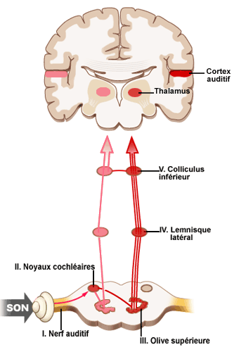
An active electrode placed on the vertex of the skull allows the recording of evoked auditory potentials from the auditory nerve and the brainstem (early potentials, waves I-V), and those of the higher auditory structures in the thalamo-cortex (late potentials). The brainstem evoked potentials (BAEPs), which have a short latency (<10 ms), are currently used clinically to test the auditory pathway up to the level of the inferior colliculus. See the diagram below for the association between the different waves making up the AEPs and their related anatomical structures.

Diagram illustrating the auditory pathway and the anatomical locations related to the the different waves of the AEP
– auditory nerve = wave I
– cochlear nuclei = wave II
– superior olive = wave III
– lateral lemniscus = wave IV
– inferior colliculus = wave V
Waves I to V make up the brainstem potentials (BAEPs).
The thalamus (medial geniculate ganglion) and the auditory cortex (temporal lobe) make up the middle and late waves (N, P) of the AEP.
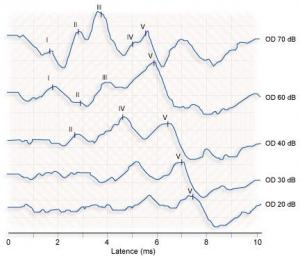
Example of recording: auditory nerve and brainstem potentials

Brainstem auditory evoked potentials (BAEPs) recorded at different sound stimulation intensities (after Legent)
This type of recording is a type of objective audiogram.
The measured potentials are weak (< µV) and so many summations may be necessary (1000 to 2000 rrepetitions) before a cleare response emerges from the background noise.
Note the presence of 5 obvious waves at 70 dB. As the intensity of the presented sound decreases, the latency of the response increases and the amplitude of the waves diminishes.
The audiometric threshold is defined as the minimum intensity needed to obtain a clear wave V, which here, as is usual in a normally hearing subject, is 20 dB.