Hair cells: overview
Authors: Rémy Pujol, Régis Nouvian, Marc Lenoir
Contributors: Sam Irving
Cochlear, as well as vestibular, sensory cells are called hair cells because they are characterised by having a cuticular plate with a tuft of stereocilia bathing in the surrounding endolymph. The cell body itself is localised in the perilymph compartment (see transverse section of the organ of Corti).
Schematically, both types of cells, inner hair cells (IHCs) and outer hair cells (OHCs), differ by their shape and the pattern of their stereocilia. Their innervation to and from the CNS is also totally different (see ‘ Organ of Corti: innervation‘).
Hair cells: structure
In the human cochlea, there are 3,500 IHCs and about 12,000 OHCs. This number is ridiculously low, when compared to the millions of photo-receptors in the retina or chemo-receptors in the nose! In addition, hair cells share with neurons an inability to proliferate they are differentiated – this means that the final number of hair cells is reached very early in development (around 10 weeks of fetal gestation); from this stage on our cochlea can only lose hair cells.
Schematic pictures of an inner (IHC) and outer (OHC) hair cell
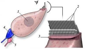
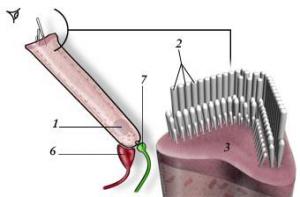
1. Nucleus, 2. Stereocilia, 3. Cuticular plate, 4. Radial afferent ending (dendrite of type I neuron), 5. Lateral efferent ending, 6. Medial efferent ending, 7. Spiral afferent ending (dendrite of type II neuron)
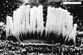
M Lenoir
Linear pattern of IHC stereocilia
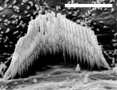
M Lenoir
“W” pattern of OHC stereocilia
In both cases 3 rows of stereocilia of graded length, linked to each other, are embedded in a glabrous(i.e. bearing no microvilli) cuticular plate (in contrast with the surface of supporting cells which bear numerous microvilli).
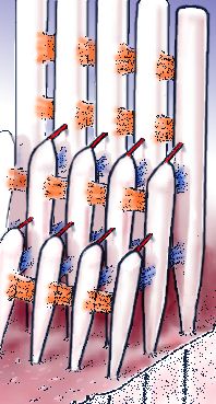
Stereocilia (around a hundred) are generally arranged in three rows of graded lengths. In addition to thin tip links (shown here in red) which are involved in the mechano-transduction process, stereocilia are attached by transverse (lateral) links, both in the same row and from row to row.
Tip (red arrow) and lateral (blue arrow) links between two stereocilia.
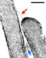
With transmission electron microscopy (TEM), the tip link (red arrow) and a lateral link (blue arrow) between medium and tall stereocilia are clearly visible. At both ends of the tip link, a membrane condensation is seen.
Electro-mechanical transduction
Hair cell stereocilia are at the core of electro-mechanical transduction; the transformation of sound vibration into a neural signal that can be interpreted by the brain. Both types of hair cell have a similar transduction mechanism.
The deflection of the stereocilia causes stretch-sensitive ion channels to open. These are non-selectively permeable to cations and are located at the base of the tip links, with 1 or 2 channels per tip link.
Stereocilia bathe in endolymph, which is rich in potassium and characterised by an endocochlear potential of +80 mV, but the hair cell body has a potential between -70 mV and -55 mV. As a result, the electric potential between the endolymph and the hair cell body (between 135 and 150 mV) causes a massive influx of potassium ions from the endolymph to the hair cell when the mechanically-sensitive ion channels open. This cation influx depolarises the hair cell.
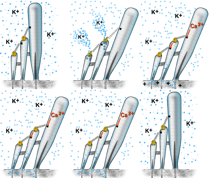
Transduction channels: opening and adaptation
Displacement of the stereocilia towards the stria vascularis causes the cation channels to open: potassium (K+) enters the hair cell, causing it to depolarise. At the same time, another cation, calcium (Ca2+), also enters the cell.
K+ channels close before the stereocilia return back to the modiolus. This is an adaptation method which allows rapid, successive, stimulation cycles to occur.
The rapid component of adaptation arises from the partial closure of the ion channel following the direct fixation of calcium to the channel. The slow component arises from the displacement of myosin along the actin filaments of the stereocilia. This causes the tip link tension to decrease and encourages the channel to close. Here also, the calcium that entered via the mechanically-sensitive ion channel is responsible for activating the myosin.