Noise induced trauma
Authors: Rémy Pujol
Contributors: Jing Wang, Sam Irving
Depending on the length of exposure, very high sound intensities (above 90 dBA) can damage the structures of the organ of Corti and cause either temporary or permanent hearing loss. At the level of the sensorineural structures (hair cells and spiral ganglion cells), noise trauma occurs at two levels: at the OHCs and at the axon terminals synapsing with the IHCs. These structures are involved to different degrees depending on the type of noise trauma, and their ability to self-repair depends upon their functional recovery.
See more information about the sounds or noise dangerous for our cochlea
Hair cell damage
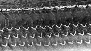
Scanning electron microscope (SEM) view of the surface of a rat cochlea
M. Lenoir
Normal cochlea
The row of IHCs and the 3 rows of OHCs have normal appearance.
Note: this image and those that follow are presented at slightly different magnifications; to get an indication of scale, the distance between the base of the ‘V’ shape OHC stereocilia is around 7 µm.
J. Wang
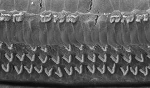
Noise trauma: stage 1
Some IHCs and some OHCs in the first row have already disappeared.
J. Wang
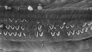
Noise trauma: stage 2
By increasing the intensity of the traumatic sound, most of the IHCs have disappeared in the frequency region corresponding to the sound. OHCs in the first row have also disappeared and damage has occurred in the 2nd and 3rd rows.
J. Wang
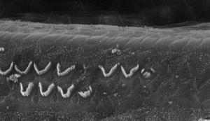
Noise trauma: stage 3
With a further increase in noise level, in the frequency region corresponding to the frequency of the sound, only a few OHCs in the 2nd and 3rd rows remain .
Duality of acoustic trauma in the cochlea: synaptic versus cell damage
Exposure to a sound over 130 dB (SPL) for 30 minutes translates into frequency-specific excitotoxic destruction of the synapses under the IHCs and the hair cells, which generally begins with the first row of the OHCs, but also involves the second row and the IHCs.
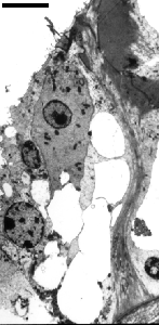
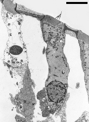
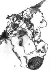
Electron microscope images of hair cells after acoustic trauma.
On the left, synaptic damage is instantaneous under the IHC. This damage can be repaired in certain cases (see excitotoxicity).
The images in the middle and to the right show OHCs a few hours after noise trauma. In the first row of OHCs (middle) or in the image on the right, the apoptotic nucleus can be seen with an advanced cytolysis. These OHCs will soon disappear.
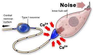
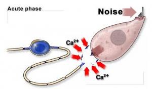
Excess noise causes an excess of glutamate to be released, causing the postsynaptic bouton to swell and burst (see excitotoxicity). This phase can be followed by neural resprouting and synaptic repair, meaning that function can be recovered (in 2-3 days).
However, in case of severe or repetitive trauma, the IHC itself can be damage (see SEM pictures above). This loss of IHC is generally followed, several days after, by an apoptotic loss of neurons, which accounts together with the HC dead for permanent hearing losses (PTS).
Temporary (TTS) versus permanent threshold shifts (PTS)
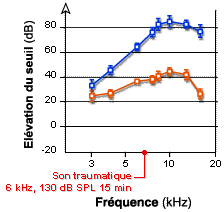
J-L. Puel, J. Ruel
Traumatic exposure (6kHz, 130 dB SPL, 15 minutes) in guinea pig immediately causes:
- Profound hearing loss (up to 80 dB, blue curve) in an unprotected cochlea
- Moderate hearing loss (up to 40 dB, red curve) in a cochlea previously perfused with kynurenate (which prevents excitotoxicity).
This indicates that, with this type of sound exposure, 50% of immediate hearing loss must be caused by synaptic damage ( excitotoxicity).