Tous droits réservés © NeurOreille (loi sur la propriété intellectuelle 85-660 du 3 juillet 1985). Ce produit ne peut être copié ou utilisé dans un but lucratif.
Soon you will find new pages dealing with research lines about:
- the cochlea, with the development of cell and molecular biology, genetics, ...
- the new methods of rehabilitation,
- the future therapies, including the next to come in situ cochlear pharmacology, but also the far-off stem cells, gene therapy, regeneration.
In the meantime, one can find some information in the general audience section " Future cures"
To open the black boxes still hiding parts of the cochlear physiology and pathology we need to develop new techniques
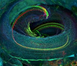
In preview: the new confocal technique of the 'cleared cochlea'
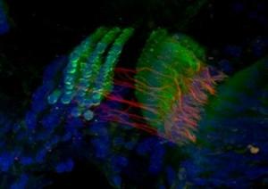
Developed in 'Rubelab' (Seattle), this technique allows a direct visualization of the whole mouse cochlea, with a confocal microscope. Among others, this should results in a much better understanding of the cellular organization of the organ of Corti. Especially, interesting informations may arise about some parts of the cochlea, which are difficult to access with classical histology: i.e., the extreme base (hook) and the extreme apex.
| G. MacDonald |
Adult mouse cochlea, after clearing of the bone and small opening in the apex. A triple immunolabeling, shows the spiral of the organ of Corti, with the hair cells (green) and the neural structures (red). All nuclei are colored in blue. |
| G. MacDonald |
High mag of the organ of Corti from the same cochlea as above. Seen clearly, the hair cells (green) and the neurites (red) reaching the base of IHCs and crossing the tunnel of Corti (probably medial efferents). All nuclei are in blue. This technic allows a 3D reconstruction and deconvolution, from optical sections. |
 Français
 English
 Español
 Português


Facebook Twitter Google+