Lateral lemniscus and inferior colliculus
Authors: Pablo Gil-Loyzaga
Contributors: Rémy Pujol, Sam Irving
The cochlear nuclei and superior olivary complex project to the higher levels of the brainstem via the lateral lemniscus. Some fibers have a relay point in the nuclei of the lateral lemniscus, whereas others project directly to neurons in the inferior colliculus.
Nuclei of the lateral lemniscus (LL)
These nuclei are composed of different neuronal groups organised between the ascending fibers of the lateral lemniscus. They are organised into two regions:
- The ventral nucleus, composed of monaural neurons that receive information from the ipsilateral ventral cochlear nucleus and are not arranged tonotopically. This nucleus has a role in processing the duration of complex sounds, which is very important for understanding language.
- The dorsal nucleus, composed of binaural neurons that recevei information from both ears, thanks to the information exchange that happens at the commissure between the lemnisci and the olivary complex.
The inferior colliculus (IC)
The inferior colliculus (IC) is situated at the roof of the mesencephalon. It is composed of two parts:
- The external and dorsal cortices, are organised in neuronal layers without precise tonotopic organisation. It has been suggested that these neurons are involved in the complex analysis of auditory information (such as language) and identification of novel sounds.
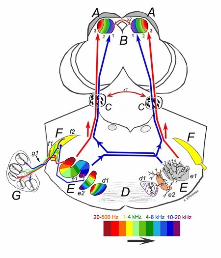
- The central nucleus is composed of a large group of tonotopically-organized neurons. This nucleus receives input from the cochlear nuclei, the superior olivary complex and the lateral lemniscus. Its structure is layered (like an onion – in colour on the figure). The frequency distribution in this nucleus is very particular, as it receives low frequency information from the ipsilateral ear, and high frequency information from the contralateral ear. Amongst other things, this nucleus exchanges information with the contralateral central IC nucleus via the commissure. This nucleus is involved in frequency analysis, but also has a role in decoding interaural intensity and time differences.
IC neurons are involved in sound localisation in the horizontal and vertical plains. Their activity is mediated by the descending fibers from the cortex and the thalamus (medial geniculate body).
Tonotopic organisation
The tonotopic organisation seen in the SOC continues in the higher nuclei of the auditory system.

P. Gil-Loyzaga
The ascending projections from the SOC pass along the lateral lemniscus (C) and arrive at the external nucleus of the inferior colliculus (A and B). The fibers that send low frequency information (in red and marked with number 3) project along the lateral side of the lemniscus (ipsilaterally) and reach the external nucleus of the IC. The fibers that send high frequency information (in blue, and number 1) cross over in the superior olivary complex and project up inside the contralateral lemniscus to the medial region of the IC. The colour scale at the bottom indicates sound frequency.