Oto-Acoustic-Emissions
Authors: Jean-Luc Puel, Pierre Bonfils, Jean Pierre Piron
Contributors: Sam Irving
Discovered back in 1978, by D. Kemp, oto-acoustic emissions (OAEs) were not fully explained until a few years later, after the OHC active mechanism had been understood. At least in the mammalian cochlea, OAEs reflect OHC electromotility. OHC contractions/elongations themselves vibrate cochlear fluids and the middle ear conducting mechanism transfers this vibration back to the air of the external auditory canal: there, the emissions can be registered by a microphone.
Schematic drawing of a probe for recording evoked OAEs
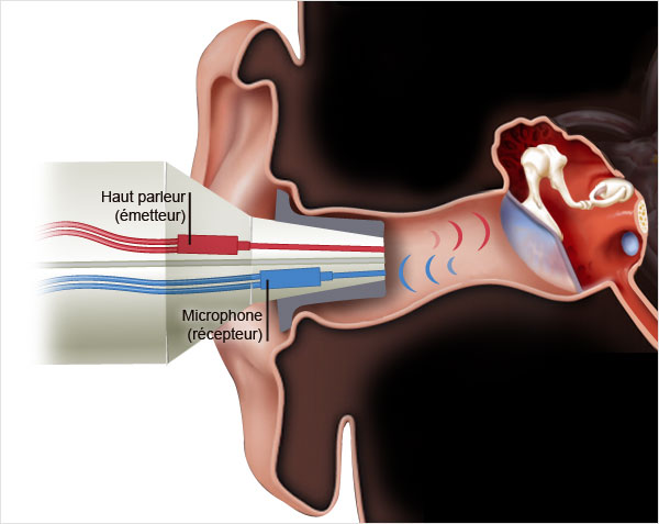
The probe contains
- 1 a speaker (red) emitting the stimulating sound
- 2 a microphone (blue) to record the returning OAEs.
Examples of evoked oto-acoustic emissions (normal subject)

OAEs in response to a click stimulation
The superimposition of two traces indicates the reproducibility of the recording. Actually yhry are remarkably constant: for the same type of stimulus in the same ear, the same OAEs will be recorded as long as the hearing is not impaired.
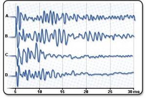
Specificity of OAEs
Each ear and each subject present characteristic OAEs in response to the same stimulus (here a click of 20 dB SPL).
- A and B are OAEs from left and right ears of a first subject.
- C and D are OAEs from left and right ears of a second subject.
Oto-acoustic emissions and audiometry
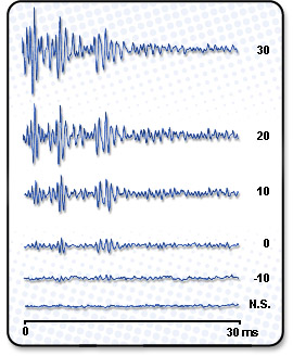
OAEs and hearing threshold
Example of evoked OAEs in response to varying intensities of stimuli.
The threshold for a normally hearing subject, as in this example, is very low: here 10 dB better than the ‘normal’ perception threshold of 0 dB! This makes OAE recording a very sensitive test.
However, the response saturates quickly: in this case at 30 dB above the perception threshold there is no further change in response.
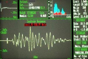
OAEs: screening and prevention
Classical recording in ENT clinic: normative data.
OAEs reflect the activity of OHCs, which are the most sensitive cells of the organ of Corti.
Thus, they can be used as an objective screening test in newborns, but also in adult subjects at risk, for example workers in noisy environments, patients undergoing therapy with ototoxic drugs.
Distorsion products
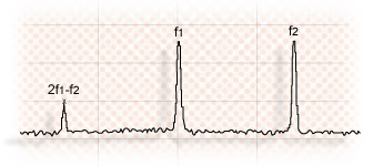
2f1-f2 distorsion products
Distortion products (DP) reflect the non linearity of a normal cochlea. In response to sound stimulation by two frequencies (f1 and f2), different DP OAEs can be recorded: the most frequently used in clinics and research is 2f1-f2.
This type of OAE is frequency specific and allows to trace a real objective audiogram, which reflects the integrity of OHCs.
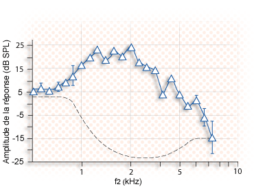
Distortion product audiogram
2f1-f2 amplitude is plotted as a function of f2 frequency.
Note a clearly identifiable response (against the noise (bottom doted line) for frequencies beween 1 and 6 kHz. Below 1 kHz, active mechanisms, if any, are not strong enough to allow any recording. Above 6 kHz, the lack of DP OAEs reflects the aquipment limits.
Spontaneous oto-acoustic emisions (SOAEs)
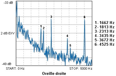
Recording of SOAEs
In about 30% of normal ears, it is possible to record spontaneaous OAEs. This means that in the absence of stimulation, a microphone in the auditory meatus may record OAEs.
These spontaneous OAEs are generally recorded at mid- or high frequencies (as the six emissions recorded in this normal hearing subject).
A precise correlation with cochlear physiology is not available. Do they reflect mild abnormalities such as missing or supernumerary OHCs?
Very rarely, the phenomenon is loud enough to be perceptible without a microphone: the ringing is heard by the subject and/or by other people.
Oto-acoustic emissions and efferent system
The medial efferent system, synapsing with OHCs, attenuates the electromotile properties of OHCs via slow contractions, thus reducing OAEs. Here are 3 examples demonstrating the phenomenon:
- a contra-lateral stimulation reduces OAEs in the opposite ear ;
- a similar effect is obtained by a direct application of acetylcholine, the main medial efferent neurotransmitter ;
- selective attention (either visual or auditory) reduces OAEs through an action of the medial efferent system driven by higher brain structures.
Thus, OAEs may be used to probe the activity of the medial efferent system.