Presbycusis
Authors: Jean-Luc Puel, Rémy Pujol
Contributors: Jing Wang, Sam Irving
Traditionally, presbycusis is defined as the ‘normal’ ageing of the ear, as opposed to an induced hearing loss caused by pathology. Hearing is the sense that ages the least gracefully: because of their comparatively small number, the disappearance of hair cells and cochlear neurons over time cause an age-related hearing loss.
Presbycusis: overview
Presbycusis is a complex condition. It has many causes, but its origin lies within a combination of individual (such as age, or genetics) and environmental (e.g. noise exposure, medication) factors. From a physiopathological point of view, it is characterised by degeneration of the organ of Corti (sensory presbycusis), and/or the spiral ganglion (neural presbycusis), and/or the stria vascularis (metabolic presbycusis).
In societies where life expectancy continues to increase, age-related hearing loss, or presbycusis, is a public health issue. In fact, 70% of the world’s population over the age of 65 have hearing problems. Symptoms are a decrease in the perception of high frequency sounds, and comprehension problems in noisy environments. With time, hearing and comprehension problems are accentuated, even in quiet environments, and can become debilitating. Alongside hearing loss, tinnitus (a ringing or whistling sound) is also common in clinical populations. Finally, age-related hearing loss can lead to social isolation which is often the cause of depressive symptoms.
Presbycusis and other hearing loss
Presbycusis is not the only type of acquired hearing loss.
The ‘natural’ ageing of the ear can be accelerated because of all of the pathologies accumulated over the years, particularly the use of ototoxic drugs (such as aminoglycoside antibiotics) and over-exposure to noise (acoustic trauma). Genetic predisposition can also cause individual variations.

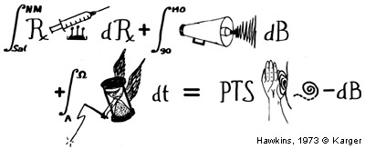
In this clever representations, Hawkins does an excellent job of characterising the degree of presbycusis by summing up the cumulative effects of ageing and the main risk factors to the cochlea. Hearing loss in dB (or PTS) is equal to the sum of ototoxic drugs and traumatic noise (over 90 dB) accumulated over the years. Nearly 40 years on, if you add in genetic effects, this ‘equation’ is still current.
“Normal” and “accelerated” presbycusis

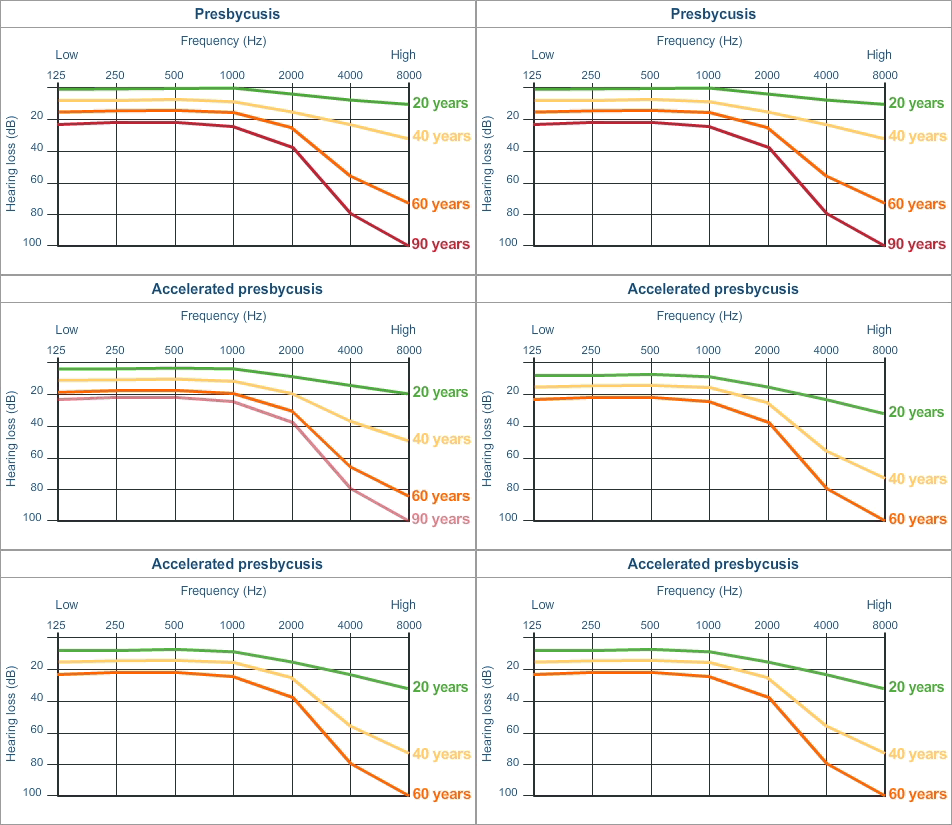
See the acceleration of presbycusis (or precocious presbycusis) caused by exposure to intense sound!
The graph above represents the average hearing levels for individuals aged 20, 40, 60 and 90 years. “Natural” ageing can be accelerated because of accumulation of pathologies over the years. For example, excessive exposure to very intense and traumatic sound can cause precocious presbycusis: at the age of 40, you can have the ears of a 90 year old!
Experimental models of presbycusis
There are a few animal models of afe-related hearing loss, amongst them the SAMP8 mouse is a good approximation of human presbycusis. Other than general ageing, these mice present with early onset hearing loss following loss of outer hair cells and damage to the strai vascularis. Perhaps more surprising is that a large loss of auditory neurons occurs from the age of six months, when the inner hair cells are still intact. In this case, the initial degeneration is in the spiral ganglion cells.
This model (and a few others) allows us to study the cellular and molecular mechanisms of degeneration in cochlear structures. Preliminary results show that cell death (apoptosis) is caused by a large inflammatory process and mitochondrial dysfunction in all of the cochlear structures. Understanding these processes enables more specific treatments for presbycusis to be developed.
Presbycusis and cortical plasticity
When the degree of presbycusis and cochlear histology can be compared in the same subject, the lack of correlation can be striking: the number of residual hair cells and/or spiral ganglion cells is often much lower than physiological testing would suggest. In other words, one can have acceptable hearing with considerable damage to regions of the cochlea. This is due mainly to a well-known phenomenon: cortical plasticity. The auditory cortex, when deprived of a certain number of afferents, manages to adapt to optimise the messages that it’s still able to receive.
For example, when the base of the cochlea deteriorates, cortical neurons that coded exclusively for high frequency sound become more responsive to mid and low frequencies, thus improving perception of these stimuli.