Ear: overview
The ear is divided in 3 parts: the external and middle ear transfer the sound waves to the inner ear, where they are transduced in neural activity.
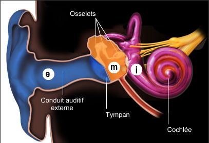
Diagram of the three parts of the ear : external ear (E), middle ear (M) and inner ear (I)

The outer or external ear (e blue) is composed of the pinna (the visible part!) and the ear canal. The latter is closed off by the eardrum. In the middle ear (m orange), the eardrum is mechanically linked by a chain of three tiny bones (the ossicles) to another membrane (the oval window) which closes the inner ear (i red). The hearing part of the inner ear is rolled up into a spiral called the cochlea, as it looks like a snail shell (‘cochlea’ is the greek word for snail).
Note. Over the cochlea we can see the vestibule, which is the second sensory organ of the inner ear. It plays a main role in the equilibrium.
This 3D animation shows the anatomical relationships between the three part of the Ear.
You can stop the animation to better figure these relationships, and compare with the above drawing.