Cochlea: overview
Authors: Guy Rebillard, Rémy Pujol
Contributors: Sam Irving
The cochlea represents the ‘hearing’ part of the inner ear and is situated in the temporal bone. It derives its name from the Greek ‘kokhliās’ (meaning ‘snail’) as it forms a spiral structure during development, which makes it resemble a snail shell.
The cochlea interacts with the middle ear via two holes that are closed by membranes: the oval window, which is located at the base of the scala vestibuli and which undergoes pressure from the stapes (see ‘middle ear’), and the round window, which seals the base of the tympanic membrane and is used to relieve pressure.
Structure of the cochlea
From the cochlea to the organ of Corti
Par zooms successifs, on observe :
- une coupe transversale de la cochlée,
- l’organe de Corti,
- la cellule ciliée, à l’origine du message nerveux, et une fibre (bleue) du nerf auditif, qui le transmet l’information au cerveau.
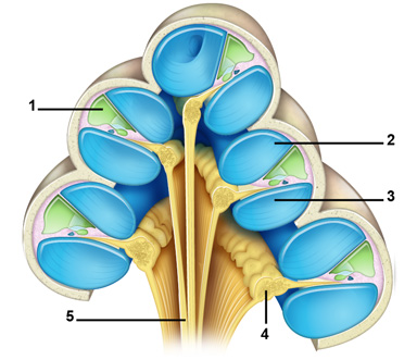

The cochlea is made up of three canals wrapped around a bony axis, the modiolus. These canals are: the scala tympani (3), the scala vestibuli (2) and the scala media (or cochlear duct) (1). The scalae tympani and vestibule are filled with perilymph (in blue) and are linked by a small opening at the apex of the cochlea called the helicotrema. The triangular scala media, situated between the scalae vestibuli and tympani is filled with endolymph (in green).
Between the scala media and the scala tympani is a structure called the organ of Corti.
The neural elements (shown in yellow) are the spiral ganglion neurons (4) and the auditory nerve (5) in the modiolar plane.
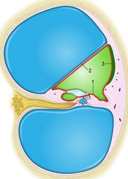
Cross-section of a single cochlear turn
The scala media, filled with endolymph (in green), is surrounded by the reticular lamina which covers the organ of Corti (1), by Reissner’s membrane (2) on the side of the scala vestibuli, and by the lateral wall (3), composed of the stria vascularis and the cochlear promontory. The organ of Corti rests on the basilar membrane.
The spiral ganglion neurones and nerve fibres are shown in yellow (see details below).
Note: In order to understand the function of the hair cells, it is important to remember that their stereocilia (‘hairs’) are bathed in endolymph, whereas the rest of the cell is in perilymph. The red line indicates the inpermeability of the endolymphatic compartment: this is due to tight junctions between cells.
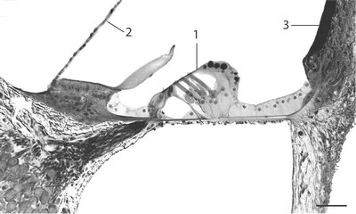
Transverse section of the 3rd turn of a rat’s cochlea, showing the Corti’s organ
During tissue preparation, the tectorial membrane has been detached from the organ of Corti and the hair cells (1). Part of the Reissner’s membrane (2) and stria vascularis (3) are indicated. The spiral ganglion neurons (bottom left) and their myelinated peripheral fibers are also clearly seen.
Scale: 50 µm
Cochlear spiral: scanning electron microscopy (SEM)
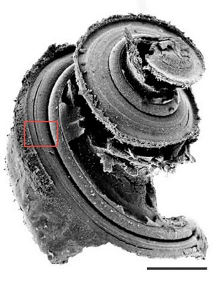
After the stria and Reissner’s membrane have been dissected from the rat cochlea, the 3 turns of the spiralling basilar membrane, supporting the organ of Corti, can be seen here at low magnification.
S.E.M. allows the visualisation of the surface of the hair cells and the organisation of their stereocilia (‘hairs’)
Frame in red points to higher magnification (below) where hair cell surfaces are seen.
Scale: 2 mm
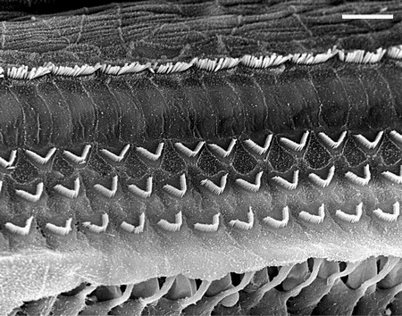
Hair cell arrangement at the base of the cochlea
Photomicrograph of the surface of the inner hair cells (top row) and the outer hair cells (3 rows below), separated by the pillars or Corti. Under the outer hair cells, a break shows the phalangeal processes of the Deiters cells below.
Scale: 15 µm