OHCs: Synapses
Authors: Rémy Pujol
Contributors: Marc Lenoir, Sam Irving
The OHCs synaptic organization correspond to their electro-motile properties. Vesiculated endings of the medial efferent system regulate the OHC motile properties. In turn the OHC synapses with small type II afferent endings, to send the brainstem, some still unclear message: perhaps a feed-back of electromotility? However, such an organization appears late during development, and it varies from base to apex in the cochlea.
Synaptic organization at the OHC base
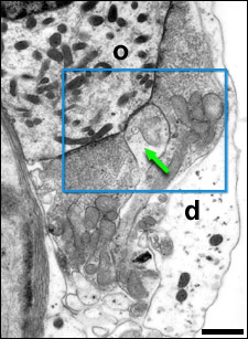
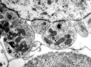
The basal pole of an OHC is contacted by large vesiculated terminals of the medial efferent system. Amongst these efferent synapses can be found one or two little dendritic boutons (green arrow) of a spiral afferent fibre (type II neurons).
This type of organisation is characteristic of most of the OHCs in that part of the cochlea that codes for the mid- and high frequencies (2 to 20 kHz).
d = Deiters’ cell; o = OHC
scale bar : 1 µm
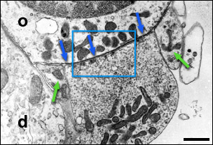

OHC in the basal turn of a rat cochlea.
A large vesiculated terminal contacts the base of an OHC (o), which is itself in close contact with the Dieters’ cell (d); green arrows indicate two small afferent dendritic boutons.
The zoom (right) allows good visualisation of the postsynaptic cistern (blue arrows) and the presynaptic microvesicles, often arranged in small packets close to the membrane.
scale bar: 1 µm
High magnification of synapses between OHC (o) and the type II afferent dendrites (a)
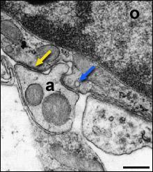
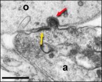
On serial sectioning, one often encounters an invagination of a spiral afferent dendritic bouton (a) in the OHC (o).
On both sides, membrane densifications (yellow arrows) can be seen. Some vesicles are seen on the presynaptic side (blue arrow).
Below, one can see the internal covering of the endocytic vesicle (red arrow). scale bars: 250 nm
scale bar: 250 nm